Objective: This experiment aims to discover if temperature has an effect on the rates of sea urchin embryonic development. By altering the external environment, the experiment will determine if temperature will affect the length of the first and subsequent cell cycles upto the pluteus larvae stage.
Introduction:
Sea urchin embryos exhibit radial holoblastic cleavage in which the first and second cleavages are both meridional and perpendicular to each other. The third cleavage is equatorial, and the fourth cleavage divides unequally to produce mesomeres, macromeres, and micromeres (Gilbert, 2003). These cleavage patterns are well understood on a group level, but it is not completely clear how the individual cells are regulated on a molecular level. One can follow the egg from fertilization to blastula to gastrula to pluteus larvae formation, but this is usually performed under ideal situations. If the conditions change, such as the salt concentration or temperature of the water, how will this affect the development of the sea urchin?
Many processes, acting in sequence or in parallel, make up the cell cycle. Many studies have shown how the cell cycle is regulated at the molecular level in the cyclic accumulation and destruction of cyclins and the cyclic activity of M phase-promoting factor (Meijer et al., 1991). However, only some of these processes affect the length of the cycle (Nurse, 1990). Other environmental factors have been suggested as additional regulators of the cell cycle. For example, temperature has been shown to have a number of effects on the role of various phases of mitotic division, such as the rate of oxygen consumption and carbon dioxide production (Hoadley, 1937). Researchers have found that cell cycle events differ greatly in their degree of temperature dependence. Sea urchins are one organism that is affected by temperature. A temperature- dependent period exists after egg fertilization in which the duration of the cleavage cycle and normal development patterns are affected by the temperature of the surrounding environment (Yamada, K, and K. Mihashi, 1998).
Sewell and Young's experiment 'Temperature limits to fertilization and early development in the tropical sea urchin Echinometra lucunter" showed that each sea urchin species has an optimal fertilization temperature based on the average temperature found in its natural habitat (Sewell, M.A.and C.M. Young, 1999). This optimal temperature is necessary for the successful development of the embryos and pluteus larvae (Katsuyuki, Y., and K. Mihashi, 1998).
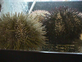
A number of species of sea urchins are found around the world. Each species thrives in their own unique environment. The subject of this experiment isLytechinus variegatus, which is found in the Gulf of Mexico and normally develops in water temperatures near 22°C.
Materials:
male and female Lytechinus variegates
100 mL beakers
sterile syringes and needles
petri dishes with covers
artificial salt water (ASW)
graduated cylinder
glass Pasteur pipets
glass depression slides
ice bucket
aluminum foil
0.5M KCl
14°C incubator
37°C incubator
thermometer
light microspcope
digital camera
Procedure:
1.Obtain gametes of sea urchins; store egg in a gently stirred suspension at 22°C, washing several times with artificial sea water (ASW)
2. Collect sperm from sea urchin and dilute one drop into 10 ml beaker containing ASW.
3. Prepare three baths of ASW at 14°C, 22°C (control), and 37° using a water bath or incubator.
4. Fertilize all the eggs with the diluted sperm in a 100 ml beaker.
5. Once fertilization envelope has developed, note time.
6. Transfer fertilized eggs and 30 ml of ASW into three glass dishes according to appropriate temperature.
7. Dilute with 30 ml of ASW at appropriate temperature (14°C, 22°C, and 38°C)
8. Cover each glass dish and place into incubator maintaining temperature.
9. Monitor the progress of development by taking a small sample and placing it on a depression slide to be viewed under a light microscope.
10. When 90% or more of the eggs have completed cytokinesis, note the time after fertilization and take still photographs.
11. Allow the fertilized embryos to develop for 24 hours.
12. After 24 hours, transfer 1 drop of each of the three samples onto depression slides.
13. Observe the sample under the light microscope. Take still photographs and compare embryonic development.
14. 48 hours after fertilization, repeat steps 12 and 13.
Results:
Temperature had an effect on the cleavage rates of developing sea urchin embryos. Generally, the higher the environmental temperature, the faster the embryo divided. At 37°C, the sea urchin eggs reached the first cell cleavage at 40 minutes after fertilization, and cleaved the second time at 65 minutes after fertilization. Abnormalities in the gastrulating embryo were observed at 37°C. Under ideal conditions, 22°C, the embryos first cleavage cycle occurred at 60 minutes after fertilization and the second cleavage cycle at 100 minutes. At 14 °C, the cleavage rate was dramatically slower, with the first division taking place at 95 minutes after fertilization. Only 50% of the embryos continued to divide, in which they reached the second cleavage at 205 minutes after fertilization. When the cultures were photographed 95 minutes after fertilization, the embryos incubated at 14°C had only divided once, the control embryos had divided twice, and the embryos incubated at 37-38°C had already divided 3 times (Figure 2).
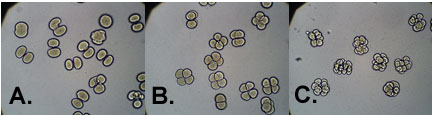
Figure 2. Sea Urchin embryos 95 minutes after fertilization. Embryos (A.) incubated at 14°C, (B.) incubated at 22°C(Control), and (C.) incubated at 38°C.
Twenty-four hours after fertilization (Day 2), the control group had developed normally. The majority of the embryos displayed archenteron formation and were motile. Not all embryos kept at 14°C had hatched by day 2. The ones that had hatched did not have archenterons and were not motile. The majority of the embryos kept at 37°C had died and lacked archenterons. The remaining embryos had developed abnormally, had not hatched and were smaller than the control group (Figure 3)
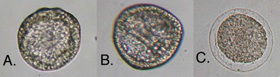
Figure 3. Sea Urchin embryos 24 hours after fertilization. Embryos (A.) incubated at 14°C, (B.) incubated at 22°C(Control), and (C.) incubated at 38°C.
Forty-eight hours after fertilization, all embryos of the control group were at the pluteus larvae stage. At 14°C, the vast majority of the embryos were dead and those alive still did not show signs of regular development such as archenteron formation. At 37°C, there were some pluteus larvae but the majority of the embryos were small and unhatched (Figure 4).
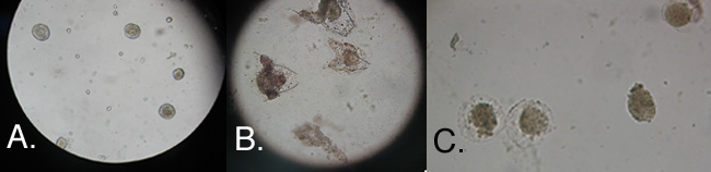
Figure 4. Sea Urchin embryos 48 hours after fertilization. Embryos (A.) incubated at 14°C, (B.) incubated at 22°C(Control), and (C.) incubated at 38°C.
Discussion:
Sea urchin development can be altered by a series of environmental changes. One of these changes is temperature. In our initial experiment, we showed that sea urchins undergo cleavage more rapidly in higher temperatures. In our second experiment observations were extended over a longer period of time. We observed that changes in temperature caused changes in development. Twenty-four hours after fertilization, archenteron formation was visible in the control embryos whereas the embryos in the other two temperatures were undergoing abnormal and slow development. Lastly, 48 hours after fertilization the control embryo were all at the pluteus larvae stage while the other embryos were either dead or developing abnormally. Overall, sea urchin embryo development is temperature- dependent, with the process occurring at a faster rate and demonstrating abnormal developments at warmer than ideal temperatures, and a slower rate, and even cell death, taking place at cooler than optimal temperatures
近日,服务科学领域的全球领导者赛默飞世尔科技(以下简称赛默飞)宣布,在达成收购意向两个月之后,赛默飞以28亿美元、折合人民币约190亿元的价格,完成了对TheBindingSiteGroup的全现金收......
11月15日,施普林格·自然和TheLens平台宣布结成重要的合作伙伴关系,以更深入地揭示学术研究和数据如何能通过经济和社会成效,加速推动创新的问题解决方式。通过将科学、投资和企业领域的开放数据更好地......
蛋白质作为构成人体组织器官的支架和主要物质,在人体生命活动中起着重要作用。蛋白质的相互作用能产生许多效应,如形成特异底物作用通道、生成新的结合位点、失活、作用底物专一性和动力学变化等,细胞的代谢、信号......
2021年9月9日,无锡臻和生物科技有限公司(以下简称“臻和科技”)与美国VyantBio公司签署TissueofOrigin®(以下简称“TOO®”)全球权益和ZL转让协议,全资收购这款唯一获FDA......
2021年7月20日,JournalofCellularPhysiology及JournalofCellularBiochemistry同时撤回了中国学者49篇文章。从2019年开始,Journalo......
6月10日,QS教育集团正式发布了2021年世界大学排名,中国共有83所高校上榜,包括内地高校51所,港澳台地区高校32所。中国大学的总体排名情况已经连续数年呈上升趋势,今年再度刷新了榜单。大学排名,......
磷酸甘油酸突变酶1(PGAM1)通过其代谢活性以及与其他蛋白质(例如α平滑肌肌动蛋白(ACTA2))的相互作用,在癌症代谢和肿瘤进展中起关键作用。变构调节被认为是发现针对PGAM1的高选择性和有效抑制......
BCEIA2019InternationalSummitonScientificInstrumentsDevelopment--RegistrationinprogressChinaisoneofth......
作为一种重要的植物激素,茉莉酸不仅调控植物对于机械损伤、昆虫取食和腐生型病原菌侵害的防御反应,还参与调控诸多生长发育过程。basicHelix-Loop-Helix(bHLH)类型转录因子MYC2是茉......
ThePlantCell是植物领域的著名学术期刊,对植物学的发展起到了重要的引领作用。为庆祝创刊30周年,ThePlantCell杂志社邀请部分编委会成员及其他科学家对发表在该杂志的重要研究工作进行评......